

Mole evaluation - Diagnosis and treatment of skin cancers
- Dermatoscopy with a portable dermatoscope for the diagnosis of “moles” and / or melanoma.
- Dermatoscopy and Mapping with the help of special software, for the monitoring of nevi (moles) and other skin lesions.
- Treatment of radial hyperkeratosis and epithelioma by various methods.
Nevus (mole) mapping in prevention of skin cancer
Skin cancers make up about 30% of malignant tumors of the human organs and systems. In 100,000 people, 90-100 suffer from skin cancer. Of all the various forms of cancer in the human body, about 4% are related to melanoma, a skin cancer that is considered one of the most malignant tumors to develop in humans. Melanoma occurs in the area of a pre-existing spilocytic nevus (mole), or in relation to it, in percentages ranging from 25% to 85%. Extensive epidemiological studies have shown that the earlier melanoma is diagnosed and treated, the more favorable its prognosis.
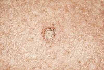
Radial keratoses (pigmented lesions that usually appear on the forehead or on the dorsal surfaces of the hands) are the most common precancerous lesions of the white skin. The probability of their transformation into squamous cell epithelium, a skin cancer that can cause metastasis, varies from 12% to 20%. Follow-up of individuals with multiple or severe radial keratoses is necessary for the appearance of new lesions and for changes in the clinical features of radial keratosis, which may indicate, with a higher probability, mutation and malignancy.
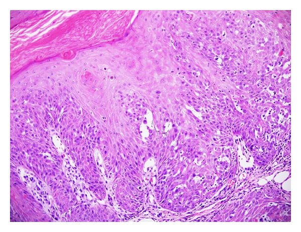
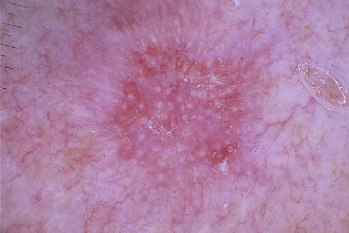
Dermatoscopy is a diagnostic technique for the examination of pigmented skin lesions or other minor lesions and rashes, which allows a more detailed and analytical observation of morphological features, that are not visible to the naked eye. It is a kind of an intermediate image between the classic clinical image and the histological one. It is applied either with a dual lens system, called a dermatoscope, and a camera, or with a digital imaging system on a computer, in order to map the moles and assess their evolution. Magnifications of these tools and machines range from 6 to 40 times, even up to 100 times. With this system, all, some or even one nevus of the skin are displayed in various magnifications, their possible malignancy is assessed, the images are stored and compared by the computer with others, which are taken at a later time. Thus, it is now possible to map skin moles and monitor their characteristics and changes over time.
Our Contacts
Contact us!
Agias Sofias 28, Thessaloniki
info@dermaesthetichair.gr
(+30) 2310 282.292 & (+30) 2310 250.300